Brodmann Areas: Characteristics And Functions
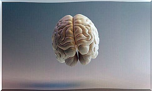
The study of the brain has always been characterized by its enormous complexity. However, some great researchers have enabled us to understand it more clearly. One of them was the German neurologist Korbinian Brodmann, who divided the cerebral cortex into Brodmann areas.
Brodmann was fundamentally dedicated to the study of anatomy and psychiatry. In 1901 he began to focus on neurobiology. It was at this time that he created the famous map of the cerebral cortex that bears his name.
On this map of Brodmann areas, we find a division of regions according to their function and location. Read on and learn more about these regions!
Brodmann areas: what do they consist of?
This map of the cerebral cortex was produced by Brodmann around 1909. There are 52 different areas there. Each of them consists of a cytoarchitectony, blood flow, metabolism and characteristic functions.
Brodmann’s goal was to create a topographical organization of the cortex, based on anatomical and functional features. To do this, he made spatial divisions of the cortex that he gradually put in relation, experimentally, with the various cortical functions.
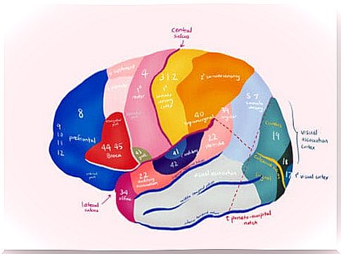
For these areas, discovered from his research in neuroanatomy, he used the Nissl staining method. Besides, he didn’t just do these studies on humans: he also did them on monkeys.
Brodmann was not the only scientist to conceive of a division of the cerebral cortex: Constantin Von Economo and Georg N. Koskinas also did, and in even more detail. However, Brodmann’s cortical map has enjoyed greater worldwide distribution and is still being used as a reference today.
Even though it is currently known that there is no exact division of cortical areas and that there are interrelationships in the cerebral cortex and not independent functions by area, this map is still of great use.
Motor areas
These are the regions which, in short, form the motor cortex. It is, therefore, a group of areas that are responsible for processing information relating to muscle movement.
They are related to the generation, coordination, maintenance and finalization of movements. We can, within the motor cortex, find different regions:
- Brodmann’s primary motor area or 4: this region is characterized by its low excitation threshold. It is responsible for executing orders to initiate voluntary movements which, in general, will be simple movements.
- Brodmann’s supplementary motor area or 6: it is characterized by a high excitation threshold. This region is responsible for coordinating the movements involved in the posture. In addition, it influences the organization of the sequences of movements of the large muscle groups.
- Brodmann’s secondary motor area or 8: this is a premotor area; with area 6, it is responsible for storing movement patterns from past experiences. She also takes care of eye movement
- Broca’s area or Brodmann’s 44 and 45: these are those linked to the movements necessary to produce language. In other words: gesticulation, intonation and semantic processing. They therefore play a crucial role in the development and generation of spoken and written language.
Brodmann areas: sensitive areas
They make up the somatosensory region and are responsible for the cerebral processing of sensory phenomena (association and coordination of stimuli and comparison of past stimuli with those arriving from outside).
They process, among other things, the information that comes from the tactile system and that relating to the body position. We can find the following sensitive regions:
- Primary somesthetic areas or 1, 2 and 3 of Brodmann: they are located between the parietal convolution and the posterior part of the central parietal lobe. These are the main areas responsible for touch and propioception
- Secondary somesthetic areas or Brodmann’s 5 and 7: 5 deals with tactile perception and 7 is an integrating area, recognizing objects without going through sight
- Regions of acoustic sensitivity: area 41 detects changes in the frequency and localization of sound; area 42 participates in the detection and recognition of speech and processes information from the primary auditory cortex. In addition, areas 20 and 21 recognize sounds and 22 perceive them.
- Taste sensitivity: this is mainly area 43. This is found in the posterior part of the lateral fissure and in the area adjacent to the insula. Thanks to it, we process the information of flavor and taste
- Vestibular sensitivity: it is found in the upper part of the central fissure and in the posterior part of the insula and of the area 22. It is linked to the perception of the positions of the body, to the movements of the head in the space and maintaining balance
- Visual sensitivity: this is Brodmann’s area 17, which is located around the calcarine fissure and the occipital pole, which processes visual content by relying on a retinotopic distribution of visual representations. In addition, Brodmann areas 18 and 19 link the information received from area 17 to recorded past visual experiences, for the recognition and appreciation of what we see.
Associative areas
They are multisensitive areas capable of associating various sensations with each other and also with motor type areas. They are related to behavior, perceptual discrimination and the interpretation of sensory experiences.
They are found in three areas: the posterior parietal, the anterior temporal and the prefrontal. So we have :
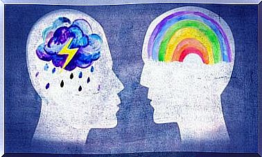
- The prefrontal areas or 9, 10, 11 and 12 of Brodmann: they are responsible for associating the experiences necessary to produce abstract ideas. They are also linked to executive functions, with personality and emotions.
- The area of the angular gyrus or Brodmann areas 39 and 40: they combine visual, propioceptive and tactile information to consolidate concepts of shape, size and texture. In addition, they are linked to the appreciation and awareness of the image.
- The anterior temporal area: it intervenes in the storage of sensitive experiences. Its stimulation makes you remember objects or music that you have experienced in the past. We attribute to area 38 of this region the movement of the foot that we make following the rhythm of a song, when we listen to a melody
- Associative language areas: Broca’s area is dedicated to the motor generation of spoken language. Wernicke’s area allows the comprehension of written and spoken language and associates sounds with concepts. The center of Exner interferes in the written language and those of Dejerine and Luria take care of the adequate organization of the word
The 97 areas of the brain that are unknown to us were identified by the HCP (Human Connectome Project) funded by the United States.